Contents
Overview
The genesis of optical microscopy can be traced to the late 16th century, with the invention of the compound microscope often attributed to Dutch spectacle makers like Zacharias Janssen and his father, Hans, around 1590. However, it was Robert Hooke)'s meticulously illustrated work, Micrographia (1665), which truly popularized the instrument and its findings, famously coining the term "cell" after observing cork. Early pioneers like Antonie van Leeuwenhoek, working independently with his own single-lens microscopes, pushed the boundaries of magnification and resolution, discovering bacteria, protozoa, and sperm cells, earning him the title "Father of Microbiology." The 17th and 18th centuries saw incremental improvements, but it wasn't until the mid-19th century that significant theoretical and practical advancements, particularly in lens design by figures like Ernst Karl Abbe, began to address the fundamental limits of resolution, paving the way for the sophisticated instruments we use today.
⚙️ How It Works
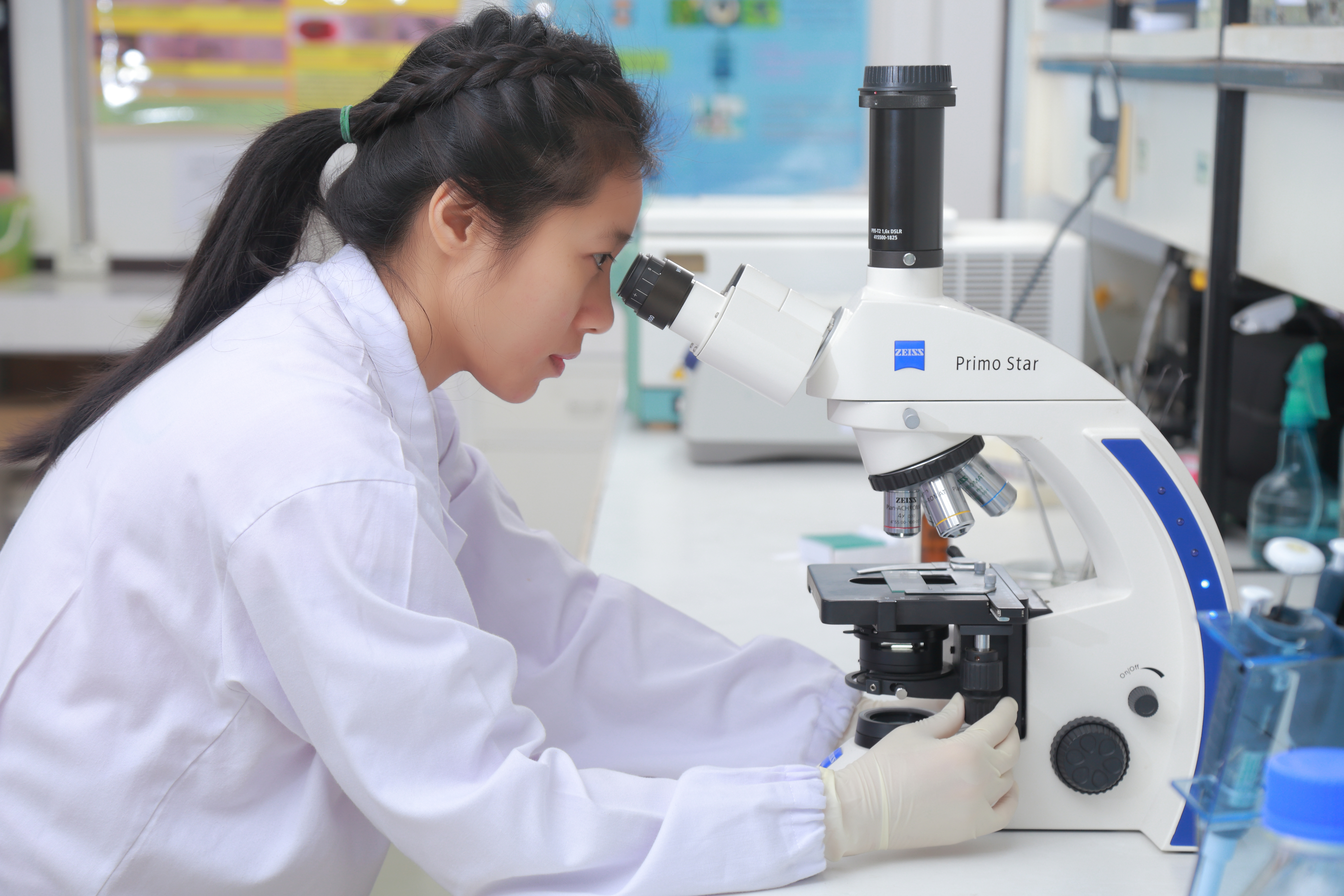
At its heart, an optical microscope functions by transmitting or reflecting light through a specimen, which is then magnified by a series of lenses. The primary components include a light source (e.g., LED or halogen lamp), a condenser to focus light onto the sample, objective lenses, mounted on a revolving nosepiece, and eyepieces (oculars) that further magnify the intermediate image. For transmitted light microscopy, samples are often thinly sliced or stained to enhance contrast. Reflected light microscopy is used for opaque specimens, with light directed onto the sample surface. Advanced techniques like phase contrast and DIC manipulate light waves to visualize unstained, transparent specimens with remarkable detail, overcoming the limitations of simple brightfield illumination. The total magnification is the product of the objective lens magnification and the eyepiece magnification, often reaching up to 1000x or more in standard configurations.
📊 Key Facts & Numbers
Optical microscopes can achieve resolutions down to approximately 200 nanometers (nm), dictated by the diffraction limit of visible light (around 400-700 nm wavelength). This resolution limit means that two objects closer than about half the wavelength of light will appear as one. Standard compound microscopes offer magnifications ranging from 40x to 1000x, with high-end research instruments capable of pushing this further. A typical laboratory microscope might cost between $500 for a basic student model and over $50,000 for an advanced research system with digital imaging capabilities. Over 1 million optical microscopes are estimated to be manufactured globally each year, serving diverse scientific and industrial sectors. The field of view, the area visible through the eyepiece, decreases as magnification increases, with a 10x objective typically yielding a field of view around 1.8 mm in diameter.
👥 Key People & Organizations
Key figures in the development of optical microscopy include Zacharias Janssen and his father, credited with early compound microscope designs around 1590. Antonie van Leeuwenhoek revolutionized observation with his superior single-lens microscopes in the late 17th century, discovering microorganisms. Ernst Karl Abbe provided the theoretical framework for resolution limits and improved lens design in the late 19th century, collaborating with Carl Zeiss and Otto Schott at Carl Zeiss AG to produce high-quality optics. Modern advancements are driven by companies like Olympus Corporation, Nikon Corporation, and Leica Microsystems, which continually innovate in areas like digital imaging, automation, and specialized microscopy techniques. The Royal Society in London has been a key platform for disseminating microscopy findings since its inception in 1660.
🌍 Cultural Impact & Influence
The cultural impact of optical microscopy is profound, fundamentally altering our understanding of life and matter. Hooke's Micrographia not only introduced the "cell" but also captivated the public imagination with detailed drawings of insects and plants, sparking widespread interest in the microscopic world. Leeuwenhoek's discoveries of "animalcules" challenged prevailing notions of spontaneous generation and laid the groundwork for germ theory. In medicine, microscopy enabled the identification of pathogens, the diagnosis of diseases like cancer, and the development of histological staining techniques. Materials science relies on it to examine grain structures, defects, and surface properties, influencing everything from metallurgy to semiconductor manufacturing. The ability to visualize the unseen has fueled scientific inquiry and inspired countless works of art and literature, cementing its place as a cornerstone of modern knowledge.
⚡ Current State & Latest Developments
Current developments in optical microscopy focus on overcoming the diffraction limit and enhancing imaging speed and specificity. Techniques like super-resolution microscopy (e.g., STORM, PALM, STED) have broken the 200 nm barrier, achieving resolutions as low as 10-20 nm by employing clever optical tricks or fluorescent probes. Confocal microscopy offers optical sectioning, allowing for 3D reconstruction of thick specimens with high contrast. The integration of artificial intelligence (AI) and machine learning is revolutionizing image analysis, automating tasks like cell counting, feature identification, and even predicting experimental outcomes. Furthermore, the development of novel fluorescent probes and genetically encoded reporters allows for real-time tracking of molecular events within living cells, a field known as live-cell imaging. Companies like EVOS and Keyence are pushing the boundaries of automation and user-friendliness in benchtop systems.
🤔 Controversies & Debates
A persistent debate in optical microscopy revolves around its inherent resolution limit, often referred to as the Abbe diffraction limit. While super-resolution techniques have made significant inroads, they often require specialized sample preparation, fluorescent labels, and can be slower or more phototoxic than conventional methods. The trade-off between resolution, speed, and sample viability remains a central challenge. Another area of contention is the interpretation of images, particularly in complex biological samples, where artifacts can be mistaken for genuine structures. The cost and complexity of advanced microscopy systems also raise questions about accessibility, creating a divide between well-funded research institutions and smaller labs or educational settings. The ongoing development of computational imaging techniques also sparks debate about the role of algorithms versus direct observation in scientific discovery.
🔮 Future Outlook & Predictions
The future of optical microscopy is poised for continued innovation, driven by the quest for higher resolution, faster imaging, and deeper biological insights. The integration of AI is expected to become even more pervasive, not only in image analysis but also in guiding experimental design and optimizing microscope parameters in real-time. Advances in nanotechnology may lead to new types of optical probes and nanostructures that can interact with light in novel ways, potentially pushing resolution even further. The development of portable, low-cost microscopy devices, perhaps leveraging smartphone technology, could democratize access to microscopic imaging in resource-limited settings and for field applications. Furthermore, the fusion of optical microscopy with other imaging modalities, such as electron microscopy or atomic force microscopy, will likely yield unprecedented multi-scale views of biological and material systems.
💡 Practical Applications
Optical microscopy is a workhorse across numerous fields. In biology and medicine, it's essential for diagnosing diseases (e.g., examining blood smears, tissue biopsies), studying cell morphology, tracking cellular processes in real-time, and developing new drugs. Materials science utilizes it for quality contro
Key Facts
- Category
- science
- Type
- topic